studybuddy.live/courses/fa23-biol-2031-human-anatomy-and-physiology-i-online/api-module-3-lab-activity-7-brain-dissection-external-brain-inferior-view?...?
le 3 Lab - Activity 7 - Brain Dissection - External brain: Inferior view
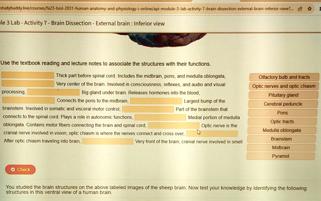
Use the textbook reading and lecture notes to associate the structures with their functions.
Thick part before spinal cord. Includes the midbrain, pons, and medulla oblongata,
Very center of the brain. Involved in consciousness, reflexes, and audio and visual
Big gland under brain. Releases hormones into the blood,
processing,
Connects the pons to the midbrain,
brainstem. Involved in somatic and visceral motor control,
connects to the spinal cord. Plays a role in autonomic functions,
oblongata. Contains motor fibers connecting the brain and spinal cord,
cranial nerve involved in vision; optic chiasm is where the nerves connect and cross over,
After optic chiasm traveling into brain,
Very front of the brain; cranial nerve involved in smell
Check
You studied the brain structures on the above labeled images of the sheep brain. Now test your knowledge by identifying the following
structures in this ventral view of a human brain.
Olfactory bulb and tracts
Optic nerves and optic chiasm
Pituitary gland
Cerebral peduncle
Pons
Optic tracts
Medulla oblongata
Brainstem
Midbrain
Pyramid
Largest bump of the
Part of the brainstem that
Medial portion of medulla
Optic nerve is the