The posterior horns contain the cell bodies of sensory neurons. These cell bodies receive input from the axons
of pseudounipolar neurons in the PNS that enter the spinal cord via the posterior root. The cell bodies of these
pseudounipolar neurons are located just lateral to the spinal cord in a swollen knob called the posterior root
ganglion.
The lateral horns are found in the thoracic and lumbar regions of the spinal cord. They contain the cell bodies
of autonomic neurons.
As you can see in Figure 13.18, it is possible to tell the anterior and posterior spinal cord apart by looking at the
shapes of the anterior and posterior horns. The anterior horns are broad and flat on the ends, whereas the posterior
horns are more tapered, and they extend farther out toward the edge.
Surrounding the spinal gray matter is the spinal white matter, which can be divided into three funiculi (fur-K-yoo-lye
also known as columns): the anterior, posterior, and lateral funiculi. Each funiculus contains myelinated axons grouped
into bundles called tracts. Tracts contain axons that have the same beginning and end points and the same general
function. Ascending tracts carry sensory information from sensory neurons to the brain, and descending tracts carry
motor information from the brain to motor neurons.
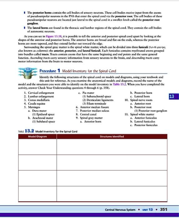
Procedure 1 Model Inventory for the Spinal Cord
Identify the following structures of the spinal cord on models and diagrams, using your textbook and
this unit for reference. As you examine the anatomical models and diagrams, record the name of the
model and the structures you were able to identify on the model inventory in Table 13.2. When you have completed the
activity, answer Check Your Understanding questions 4 through 6 (p. 358).
1. Cervical enlargement
2. Lumbar enlargement
3. Conus medullaris
4. Cauda equina
5. Meninges
a. Dura mater
(1) Epidural space
b. Arachnoid mater
(1) Subdural space
c. Pia mater
(1) Subarachnoid space
(2) Denticulate ligaments
(3) Filum terminale
6. Anterior median fissure
7. Posterior median sulcus
8. Central canal
9. Spinal gray matter
a. Anterior horn
TABLE 13.2 Model Inventory for the Spinal Cord
Model/Diagram
Structures Identified
b. Posterior horn
c. Lateral horn
10. Spinal nerve roots
a. Anterior root
b. Posterior root
(1) Posterior root ganglion
11. Spinal white matter
a. Anterior funiculus
b. Lateral funiculus
c. Posterior funiculus
Central Nervous System UNIT 13 351
13