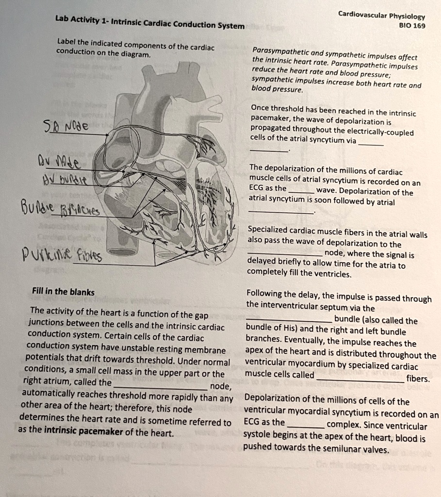
Label the indicated components of the cardiac conduction on the diagram.
Fill in the blanks:
The activity of the heart is a function of the gap junctions between the cells and the intrinsic cardiac conduction system. Certain cells of the cardiac conduction system have unstable resting membrane potentials that drift towards threshold. Under normal conditions, a small cell mass in the upper part of the right atrium, called the SA node, automatically reaches threshold more rapidly than any other area of the heart; therefore, this node determines the heart rate and is sometimes referred to as the intrinsic pacemaker of the heart.
Parasympathetic and sympathetic impulses affect the intrinsic heart rate. Parasympathetic impulses reduce the heart rate and blood pressure; sympathetic impulses increase both heart rate and blood pressure.
Once threshold has been reached in the intrinsic pacemaker, the wave of depolarization is propagated throughout the electrically-coupled cells of the atrial syncytium via the AV node.
The depolarization of the millions of cardiac muscle cells of the atrial syncytium is recorded on an ECG as the P wave. Depolarization of the atrial syncytium is soon followed by atrial contraction.
Specialized cardiac muscle fibers in the atrial walls also pass the wave of depolarization to the AV node, where the signal is delayed briefly to allow time for the atria to completely fill the ventricles.
Following the delay, the impulse is passed through the interventricular septum via the bundle of His and the right and left bundle branches. Eventually, the impulse reaches the apex of the heart and is distributed throughout the ventricular myocardium by specialized cardiac muscle cells called Purkinje fibers.
Depolarization of the millions of cells of the ventricular myocardial syncytium is recorded on an ECG as the QRS complex. Since ventricular systole begins at the apex of the heart, blood is pushed towards the semilunar valves.