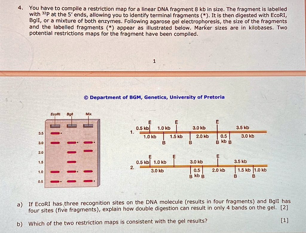
4. You have to compile a restriction map for a linear DNA fragment 8 kb in size. The fragment is labelled
with 32P at the 5' ends, allowing you to identify terminal fragments (*). It is then digested with EcoRI,
BglI, or a mixture of both enzymes. Following agarose gel electrophoresis, the size of the fragments
and the labelled fragments (*) appear as illustrated below. Marker sizes are in kilobases. Two
potential restrictions maps for the fragment have been compiled.
3.5
3.0
2.0
1.5
1.0
0.5
1
Department of BGM, Genetics, University of Pretoria
EcoRI BglI Mix
E
E
0.5 kb 1.0 kb
1.
1.0 kb
E
1.5 kb
3.0 kb
2.0 kb
B
B
0.5
B kb B
3.5 kb
3.0 kb
E
E
E
0.5 kb 1.0 kb
2.
3.0 kb
3.0 kb
0.5
B kb B
3.5 kb
2.0 kb
1.5 kb 1.0 kb
B
B
a) If EcoRI has three recognition sites on the DNA molecule (results in four fragments) and BglI has
four sites (five fragments), explain how double digestion can result in only 4 bands on the gel. [2]
b) Which of the two restriction maps is consistent with the gel results?
[1]