<Ch 03 HW
Foundation Figure 3.2: Observing Microorganisms Through a Microscope
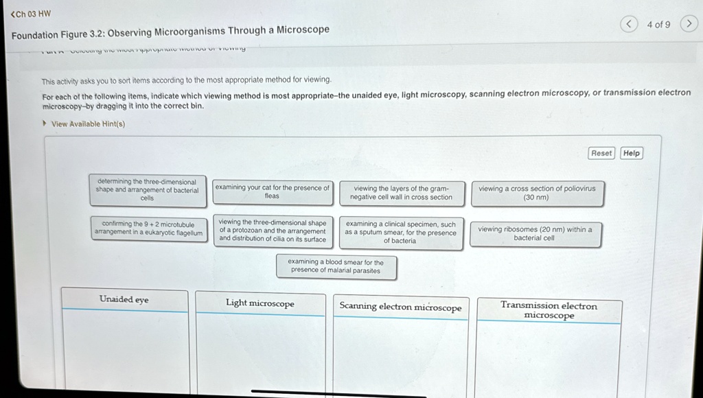
This activity asks you to sort items according to the most appropriate method for viewing.
For each of the following items, indicate which viewing method is most appropriate-the unaided eye, light microscopy, scanning electron microscopy, or transmission electron
microscopy-by dragging it into the correct bin.
► View Available Hint(s)
Reset Help
determining the three-dimensional
shape and arrangement of bacterial
cells
examining your cat for the presence of
fleas
viewing the layers of the gram-
negative cell wall in cross section.
viewing a cross section of poliovirus
(30 nm)
confirming the 9 + 2 microtubule
arrangement in a eukaryotic flagellum
viewing the three-dimensional shape
of a protozoan and the arrangement
and distribution of cilia on its surface
examining a clinical specimen, such
as a sputum smear, for the presence
of bacteria
examining a blood smear for the
presence of malarial parasites
viewing ribosomes (20 nm) within a
bacterial cell
Unaided eye
Light microscope
Scanning electron microscope
Transmission electron
microscope