Procedure 2 Brain Dissection
Often structures of the brain and spinal cord are difficult to see on anatomical models. This exercise will allow
you to examine these structures more closely by dissecting a preserved sheep brain. You will note that certain
ructures, such as the frontal lobes of the cerebral hemispheres, are proportionally smaller in the sheep than in the human
rain. Note that the process of preservation makes many structures of the brain much tougher than they would be in a
resh specimen.
1 If the brain is still encased in the skull, you have your work cut out for you. The
best way to approach extracting it from the skull is to take a hammer and chisel
and gently (at least as gently as one can with a hammer and chisel) remove it piece
by piece.
Safety Note
Goggles and gloves
are required!
2 As you remove the skull, you will note a thick membrane holding the skull in place. This is the dura mater, and it
can make removal of the skull somewhat difficult. Ideally, you would like to preserve the dura, but you may end up
cutting through it as you remove the brain.
3 Once you have removed most of the skull, gently lift out the brain. (If you're careful, you may be able to get the
brain out with the pituitary gland still attached.) You may have to loosen the remaining attachments of the dura
with your finger.
4 Once the brain is out, note the thick part of the dura covering the longitudinal fissure. If you cut through this with
scissors, you will enter the superior sagittal sinus.
5 Next remove the dura to reveal the thin membrane on top of the brain. This is the arachnoid mater.
6 Remove an area of the arachnoid mater to see the shiny inner membrane-the pia mater-directly touching the
surface of the brain. Note that the pia mater follows the convolutions of the gyri and sulci.
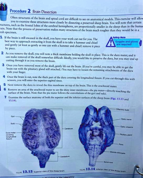
7 Examine the surface anatomy of both the superior and the inferior surfaces of the sheep brain (Figs. 13.13 and
13.14).