al Article
C. Genome
organization in vivo
1) 1000-fold
condensation
2) Spatial
organization
Nucleoid Cell membrane
B. Random coil
of the DNA
oriC
oriC
L
R
um
dif
dif
0.5 µm
Random coil volume = ~523 µm³
Nucleoid volume < 1.0 µm³
A. Circular E. coli genome
oriC
Ori
NS-L
NS-R
Escherichia coli
genome
4,641,652 bp
Left
Right
240 µm
Ter
O
dif
Inherent
polymeric
property
-Journal Article
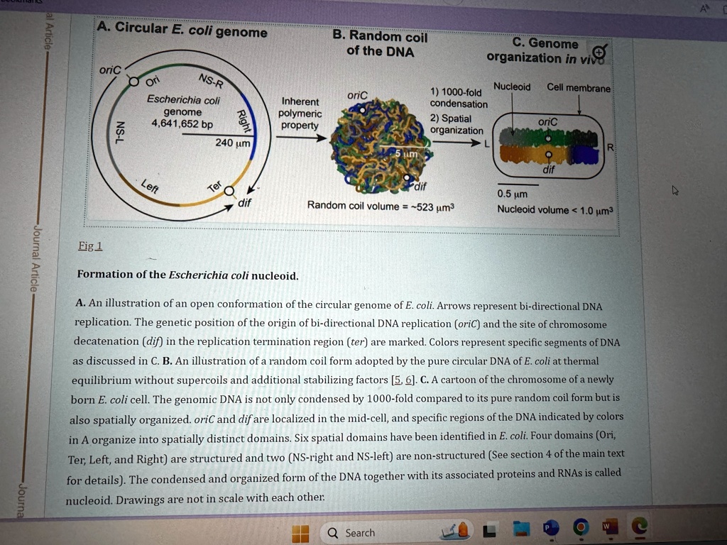
Fig 1
Formation of the Escherichia coli nucleoid.
A. An illustration of an open conformation of the circular genome of E. coli. Arrows represent bi-directional DNA
replication. The genetic position of the origin of bi-directional DNA replication (oriC) and the site of chromosome
decatenation (dif) in the replication termination region (ter) are marked. Colors represent specific segments of DNA
as discussed in C. B. An illustration of a random coil form adopted by the pure circular DNA of E. coli at thermal
equilibrium without supercoils and additional stabilizing factors [5, 6]. C. A cartoon of the chromosome of a newly
born E. coli cell. The genomic DNA is not only condensed by 1000-fold compared to its pure random coil form but is
also spatially organized. oriC and dif are localized in the mid-cell, and specific regions of the DNA indicated by colors
in A organize into spatially distinct domains. Six spatial domains have been identified in E. coli. Four domains (Ori,
Ter, Left, and Right) are structured and two (NS-right and NS-left) are non-structured (See section 4 of the main text
for details). The condensed and organized form of the DNA together with its associated proteins and RNAs is called
nucleoid. Drawings are not in scale with each other.
Q Search
e
Journa